Spinal Anatomy⁚ Modern Concepts
This book provides a comprehensive overview of spinal anatomy, including its dynamic nature, minimally invasive surgical techniques, and biomechanics. It offers a modern perspective on the field, highlighting the latest advancements in research and clinical practice.
Introduction
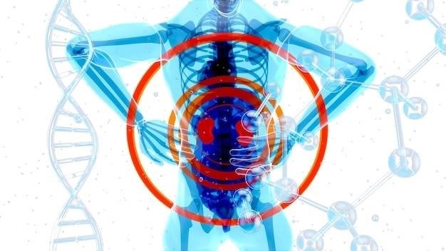
The spine, or vertebral column, is a complex and intricate structure that serves as the central axis of the human body. It comprises a series of interconnected bones called vertebrae, separated by intervertebral discs and supported by ligaments and muscles. This intricate arrangement provides structural support, protects the spinal cord and nerves, and facilitates movement and flexibility. Spinal anatomy has been a subject of intense study for centuries, but recent advancements in imaging technologies, surgical techniques, and biomechanical understanding have revolutionized our comprehension of this vital structure. This book delves into the modern concepts of spinal anatomy, exploring the dynamic nature of the spine, the impact of minimally invasive spine surgery (MISS), and the intricate biomechanics governing its function.
The Importance of Spinal Anatomy
A thorough understanding of spinal anatomy is paramount for healthcare professionals involved in the diagnosis, treatment, and management of spinal disorders. Knowledge of the spine’s intricate structure, including its bones, discs, ligaments, muscles, and nerve supply, is essential for accurately identifying and interpreting clinical presentations. This knowledge is crucial for guiding diagnostic imaging and procedures, such as magnetic resonance imaging (MRI) and computed tomography (CT) scans. Furthermore, a comprehensive grasp of spinal anatomy underpins the development of effective surgical interventions, ensuring precise placement of implants, minimizing damage to surrounding tissues, and maximizing postoperative outcomes. In essence, a deep understanding of spinal anatomy serves as the foundation for informed decision-making in the field of spine care, leading to improved patient outcomes and enhanced quality of life.
Key Structures of the Spine
The spine, or vertebral column, is a complex and vital structure composed of several key elements that work in harmony to provide support, flexibility, and protection. The foundation of the spine is comprised of vertebrae, individual bony units stacked upon one another. These vertebrae are interconnected by intervertebral discs, which act as shock absorbers and permit movement between adjacent vertebrae. The intricate network of ligaments and muscles surrounding the spine provides stability and allows for controlled movement. Finally, the spinal cord, a vital bundle of nerves extending from the brain, runs through the central canal of the vertebrae, transmitting signals throughout the body. Understanding the interplay of these structures is crucial for comprehending the biomechanics of the spine and for diagnosing and treating spinal conditions.
Vertebrae
Vertebrae, the building blocks of the spine, are individual bones that stack upon one another to form the vertebral column. Each vertebra consists of a body, a ring-like structure called the vertebral arch, and several bony projections. The body is the main weight-bearing part of the vertebra, while the vertebral arch encloses the spinal canal, protecting the delicate spinal cord. The bony projections, known as processes, serve as attachment points for muscles and ligaments, contributing to the spine’s stability and movement. There are 33 vertebrae in the human spine, grouped into five distinct regions⁚ cervical (neck), thoracic (chest), lumbar (lower back), sacral (pelvis), and coccygeal (tailbone). These regions differ in size, shape, and orientation, reflecting their specific functions. The cervical vertebrae are small and mobile, allowing for head movement, while the lumbar vertebrae are larger and more robust, supporting the weight of the upper body. The sacral vertebrae are fused into a single bone, the sacrum, which connects the spine to the pelvis.
Intervertebral Discs
Intervertebral discs are resilient, shock-absorbing structures positioned between adjacent vertebrae. Composed of a tough outer layer called the annulus fibrosus and a soft, gel-like center known as the nucleus pulposus, these discs act as cushions, enabling the spine to flex, extend, and rotate. The annulus fibrosus, composed of concentric layers of collagen fibers, provides strength and stability to the disc. The nucleus pulposus, rich in water and proteoglycans, acts as a shock absorber, distributing forces evenly across the disc. As we age, the discs naturally lose water content, leading to a decrease in their cushioning ability and increased susceptibility to injury. Degenerative disc disease, a common condition that affects the intervertebral discs, can cause pain, stiffness, and reduced mobility. Understanding the structure and function of intervertebral discs is crucial for diagnosing and managing spinal disorders.
Ligaments and Muscles
The spine’s intricate network of ligaments and muscles plays a vital role in maintaining its stability, flexibility, and proper function. Ligaments, strong fibrous bands, connect bones together, providing support and limiting excessive movement. The most important ligaments in the spine include the anterior and posterior longitudinal ligaments, which run along the front and back of the vertebral bodies, and the interspinous and supraspinous ligaments, which connect the spinous processes of the vertebrae. Muscles, attached to bones via tendons, allow for movement and provide further stabilization. The erector spinae muscles, a group of muscles running along the back, are responsible for extending the spine and maintaining posture. Other important muscles include the quadratus lumborum, which helps with lateral flexion, and the abdominal muscles, which provide core strength and support. Understanding the intricate interplay of ligaments and muscles is essential for comprehending the biomechanics of the spine and for effectively treating spinal conditions.
Spinal Cord and Nerves
The spinal cord, a vital part of the central nervous system, extends from the brain stem down the vertebral canal, protected by the vertebrae. It serves as the communication pathway between the brain and the rest of the body, transmitting motor commands and sensory information; The spinal cord is composed of gray matter, containing nerve cell bodies, and white matter, containing nerve fibers. Nerves branch off the spinal cord at each vertebral level, forming the peripheral nervous system, which innervates the entire body. These nerves control movement, sensation, and autonomic functions like breathing and digestion. The spinal cord is divided into 31 segments, each associated with a pair of spinal nerves. Understanding the anatomy of the spinal cord and nerves is crucial for diagnosing and treating spinal conditions that affect the nervous system.
Modern Concepts in Spinal Anatomy
Modern concepts in spinal anatomy emphasize a dynamic and functional approach, recognizing the spine’s intricate interplay of bones, discs, ligaments, muscles, and nerves. This understanding has revolutionized our understanding of spinal pathologies and treatment strategies. The advent of minimally invasive spine surgery (MISS) exemplifies this shift, prioritizing less invasive techniques for treating spinal disorders while minimizing disruption to surrounding tissues. MISS aims to preserve the spine’s natural biomechanics, allowing for faster recovery and improved outcomes. Furthermore, advancements in imaging technologies and biomechanical modeling have provided valuable insights into the complex interactions within the spine, enabling more precise diagnoses and personalized treatment plans. These modern concepts are crucial for improving patient care, promoting recovery, and advancing the field of spinal surgery.
Dynamic Anatomy
Dynamic anatomy recognizes the spine’s intricate movement and adaptability, challenging the traditional static view of the vertebral column. This perspective considers the complex interplay of muscles, ligaments, and joints that allow for a wide range of motions, from simple bending and twisting to complex movements during walking and lifting. Understanding dynamic anatomy is crucial for comprehending the biomechanics of the spine and its role in maintaining posture, stability, and movement. This concept highlights the importance of considering the spine’s functional capacity in both health and disease, particularly when diagnosing and treating spinal conditions. By analyzing the spine’s dynamic behavior, clinicians can identify subtle changes in movement patterns, muscle imbalances, and joint restrictions, leading to more accurate diagnoses and tailored treatment plans.
Minimally Invasive Spine Surgery (MISS)
Minimally invasive spine surgery (MISS) represents a paradigm shift in spinal surgery, emphasizing less invasive approaches to address spinal conditions. This innovative technique utilizes smaller incisions, specialized instruments, and advanced imaging technology to minimize tissue damage and reduce post-operative pain and recovery time. MISS aims to preserve the integrity of surrounding muscles, ligaments, and nerves, promoting faster rehabilitation and a quicker return to functional activities. This approach offers several advantages over traditional open surgery, including reduced blood loss, shorter hospital stays, and a lower risk of complications. The use of minimally invasive techniques has revolutionized the treatment of various spinal disorders, from disc herniations and spinal stenosis to spinal tumors and deformities. MISS continues to evolve with advancements in surgical instrumentation, imaging technology, and surgical techniques, offering promising solutions for patients seeking less invasive and more effective treatment options for their spinal conditions.
Biomechanics of the Spine
The biomechanics of the spine encompass the intricate interplay of forces, movements, and structures that govern its stability, flexibility, and load-bearing capacity. Understanding these principles is crucial for comprehending the mechanisms behind spinal disorders, guiding treatment strategies, and developing innovative interventions. The spine’s biomechanical properties are influenced by the complex arrangement of vertebrae, intervertebral discs, ligaments, and muscles. These structures work in concert to provide support, allow for movement, and absorb forces during activities like walking, lifting, and bending. The biomechanical principles governing spinal function include the distribution of forces, the role of joint mechanics, and the interplay of muscle activation patterns. These concepts are fundamental for analyzing spinal posture, identifying biomechanical risk factors for spinal disorders, and designing effective treatments for various conditions, ranging from back pain to spinal deformities.
Clinical Applications of Spinal Anatomy
A thorough understanding of spinal anatomy is paramount in the diagnosis, treatment, and management of a wide spectrum of spinal conditions. This knowledge forms the foundation for accurate assessments, effective interventions, and personalized care plans. Clinical applications of spinal anatomy encompass a range of diagnostic procedures, treatment modalities, and research endeavors. By comprehending the intricate anatomy of the spine, healthcare professionals can effectively diagnose spinal disorders, such as herniated discs, spinal stenosis, and scoliosis, and tailor treatment plans based on the specific anatomical structures involved. This knowledge also informs surgical techniques, guiding minimally invasive procedures and ensuring optimal outcomes. Furthermore, research in spinal anatomy contributes to the development of new treatments, technologies, and diagnostic tools, ultimately enhancing the quality of care for patients with spinal conditions. The clinical applications of spinal anatomy underscore its crucial role in advancing the field of spinal healthcare, fostering improved patient outcomes and a deeper understanding of the complexities of the human spine.
Diagnosis of Spinal Conditions
A precise understanding of spinal anatomy is essential for accurately diagnosing spinal conditions. Clinicians rely on a combination of physical examinations, imaging studies, and neurophysiological tests to pinpoint the source of spinal pain and dysfunction. Physical examinations involve assessing posture, range of motion, muscle strength, and reflexes, providing insights into the potential involvement of specific anatomical structures. Imaging studies, such as X-rays, CT scans, and MRIs, offer detailed visual representations of the spine, revealing abnormalities such as disc herniations, spinal stenosis, and fractures. Neurophysiological tests, including nerve conduction studies and electromyography, evaluate the function of nerves and muscles, helping to identify nerve compression or damage. By integrating these diagnostic tools with a comprehensive understanding of spinal anatomy, healthcare professionals can formulate accurate diagnoses, guiding appropriate treatment strategies and improving patient outcomes.
Treatment of Spinal Disorders
The treatment of spinal disorders is guided by a thorough understanding of spinal anatomy; Treatment options range from conservative approaches to surgical interventions, tailored to the specific condition and individual patient needs. Conservative treatments often include physical therapy, medication, and lifestyle modifications. Physical therapy aims to strengthen muscles, improve flexibility, and reduce pain, while medication can alleviate pain and inflammation. Lifestyle modifications, such as weight management and ergonomic adjustments, help to minimize strain on the spine. In cases where conservative treatments fail, surgical interventions may be considered. Surgical procedures, such as spinal fusion, discectomy, and decompression, are designed to correct anatomical abnormalities, relieve pressure on nerves, and stabilize the spine. The choice of treatment approach is based on a comprehensive evaluation of the patient’s condition, taking into account anatomical considerations, functional limitations, and individual preferences.
Research and Innovation
Research and innovation in spinal anatomy are constantly evolving, driven by the need to improve diagnosis, treatment, and management of spinal disorders. Advanced imaging techniques, such as magnetic resonance imaging (MRI) and computed tomography (CT), provide detailed anatomical insights, enabling more precise diagnosis and treatment planning. Biomechanical studies using sophisticated computer models and cadaveric specimens are shedding light on the forces and stresses acting on the spine during various activities, contributing to the development of innovative surgical techniques and prosthetic devices. Researchers are also exploring regenerative medicine approaches, such as stem cell therapy and tissue engineering, aiming to regenerate damaged spinal tissues and promote healing. These innovations hold promise for improving patient outcomes and enhancing the quality of life for individuals with spinal conditions.
Understanding spinal anatomy is paramount for effective diagnosis, treatment, and management of spinal disorders. Modern concepts in spinal anatomy emphasize the dynamic nature of the spine, the importance of biomechanics, and the evolving role of minimally invasive surgical techniques. Ongoing research and innovation are constantly refining our understanding of the intricate workings of the spine, leading to more precise diagnostic tools, minimally invasive treatment options, and innovative regenerative medicine approaches. The future of spinal care holds immense promise, with continued advancements in technology and a deeper understanding of the complex interplay of anatomy, biomechanics, and pathology.